You’re working late on a deadline when someone emails about an urgent meeting tomorrow. Within seconds, your body prepares you for action and your cortisol levels rise.
Cortisol isn’t inherently problematic; it’s a sophisticated chemical messenger that helped our ancestors survive countless threats. Yet in today’s environment this evolutionary advantage has become a potential liability.
Beyond the anxiety and sleep disruption, research connects chronic cortisol elevation to memory impairment, immune suppression, and a bunch of metabolic problems. Understanding this connection can help you better manage your cortisol levels to support both your mental and physical wellbeing.
When Protection Becomes Poison
To understand why stress affects us so profoundly, we need to look at how cortisol actually works in the body. The body’s response to stress follows a precise neurobiological pathway.
When the brain perceives a threat – psychological or physical – the hypothalamus activates the HPA axis. This triggers a series of hormonal signals culminating in the adrenal glands releasing cortisol into the bloodstream.
This mechanism is a sophisticated adaptation where cortisol serves several vital functions during acute stress:
- Releases glucose for immediate energy needs
- Boosts brain signaling for sharper thinking
- Upregulates receptor sites, making neurons more responsive
- Temporarily redirects energy away from “non-essential” systems
- Modulates immune responses to prioritize immediate survival
This is your brain on cortisol. Then, when the threat passes, baseline cortisol levels are restored and the body returns to homeostasis. These changes are remarkably effective for managing short-term challenges, the acute stressors our physiological systems evolved to handle.
The problems arise not from the cortisol response itself, but from its persistent activation. Our physiology isn’t designed for the prolonged stress that characterizes modern life: the toxic productivity, financial instability, information overload, and social complexity.
Chronic elevation of cortisol creates serious biological consequences: memory and executive functions decline, neuronal structures change, and widespread metabolic problems emerge. These disruptions lead to both mental and physical health challenges.
The good news is that our cortisol response isn’t fixed – it can be modulated through intentional practices. This adaptability gives you an opportunity to improve your cortisol regulation.
3 Simple Ways to Manage Your Cortisol Levels
While we can’t eliminate stress entirely, we can develop habits that help our bodies process it more effectively. The following strategies can make a significant difference in your cortisol levels, often within just a few weeks of consistent practice:

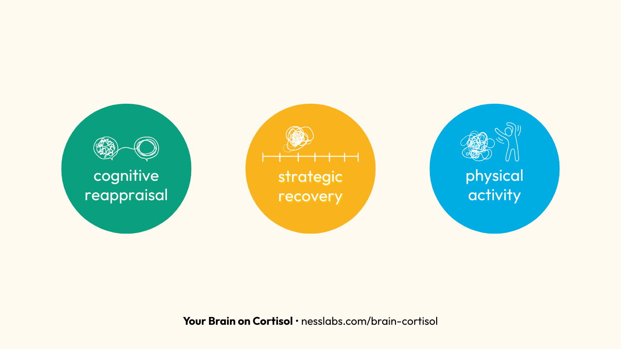
1. Cognitive reappraisal. How you think about stress actually changes how it affects your body. When you feel your heart racing, try reframing it like this: “My body is helping me rise to this challenge.” This mental shift doesn’t just feel better – research shows it actually changes how your cardiovascular system responds.
2. Strategic recovery. Working non-stop keeps cortisol levels consistently elevated. Instead, try working in focused 60-90 minute blocks followed by short breaks. During these recovery periods, take a brief walk, practice deep breathing, or chat with a colleague. This works with your body’s natural cortisol rhythms instead of fighting against them.
3. Physical activity. Exercise is a powerful cortisol regulator, but intensity matters. Moderate, consistent activity (30-40 minutes, 3-5 times weekly) can help but excessive high-intensity workouts can actually worsen cortisol imbalances. Activities with rhythmic movements – walking, swimming, cycling – are particularly effective at regulating cortisol.
You don’t need to completely eliminate stress – that’s neither possible nor desirable. Instead, aim for a healthier relationship with stress where cortisol serves its proper function without staying elevated for too long.
The most effective approach is personal experimentation. Conduct tiny experiments based on the strategies above for a few weeks, notice which ones make you feel better, and build them into your routine.
And remember: our bodies possess remarkable adaptability when it comes to stress. While evolution created our cortisol response system, your daily habits and thought patterns continuously reshape it.
